This summer, I worked on some science that was totally different from what I’m used to. Rather than tracking seed germination or collecting data on plants given different herbicide treatments in the greenhouse, I was cultivating and maintaining a diverse array of microorganisms in the lab, visualizing them under the microscope, and characterizing their physical appearance, metabolisms, and genetics. This change was brought upon by the Microbial Diversity Course (MD 2022), which I was extremely fortunate to be a part of for the last six weeks at the Marine Biological Laboratory in Woods Hole, MA. Originally, I applied to this course because I wanted to gain more microbiology experience that I could then apply to my PhD research on plant-bacteria interactions. I did not expect, however, to become completely enamored with the microbial world. I learned so much not only from the course material but also from the brilliant people I interacted with, such as the course directors, Rachel Whitaker (University of Illinois) and Scott Dawson (UC Davis), teaching and course assistants, and, of course, the other 19 scientists who were students alongside me. Here are some highlights from what I learned during this unique experience at ‘microbiology bootcamp.’
Microbes can influence our environment in obvious ways
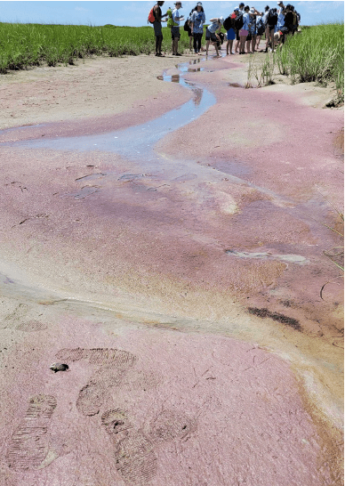

In the first week of the course, we visited the Great Sippewissett Marsh, a large tidal salt marsh near Woods Hole in Falmouth, MA, to collect samples that we would later use to cultivate microbes from. In one part of the marsh, you could see long stretches of eye-catching pinkish-purple sand. As I learned from environmental microbiologist Elizabeth Wilbanks (UC Santa Barbara) who specializes in studying microbes from this region, what gives the sand this beautiful color are microbes called purple sulfur bacteria. They are so-called because of their purple pigment and their ability to metabolize sulfur, which is highly abundant in this ecosystem. In other parts of the marsh, you can also find ‘microbial mats’, which look like thin leather rugs on the sand. If you dig up an inch or two of a microbial mat, you will see an obvious gradient of colors that are also caused by microbes. The top brownish-green leathery layer is composed of cyanobacteria. It’s followed by a pinkish-purple layer of purple sulfur bacteria, a greenish-grey layer of green sulfur bacteria, and black and grey layers of iron sulfide, the product of microbial sulfur metabolism.
To know it is to grow it
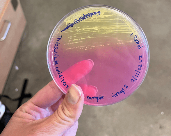
We performed a total of 24 ‘enrichments’ (techniques to enhance the growth of a particular type of microbe from a sample) to study different kinds of microbes in the lab. One of the coolest groups of microbes we grew were bioluminescent or light-emitting bacteria. These are usually members of the Vibrio and Photobacterium genus and are surprisingly easy to cultivate from seawater, sediments, and surfaces of fish and other marine life. In general, the purpose of bioluminescence for marine bacteria is unclear, but it’s known that some produce light as a result of a symbiosis with marine animals: the animals provide nutrients and habitat to the bacteria in exchange for defense, prey attraction, or other services. Bobtail squids are a cool example of an animal that participates in this special symbiosis. Other interesting microbes that we enriched for were spirochaetes – spiral-shaped bacteria that swim quickly in a corkscrew-like motion, thiosulfate-oxidizing bacteria – bacteria that live in oxygen-sulfur gradients and can oxidize thiosulfate (S2O32-) in their metabolism, microbial eukaryotes such as amoeba and ciliates (unicellular organisms with hairlike structures called cilia that help them swim and capture food), and bacteriaophages – viruses that infect bacteria.

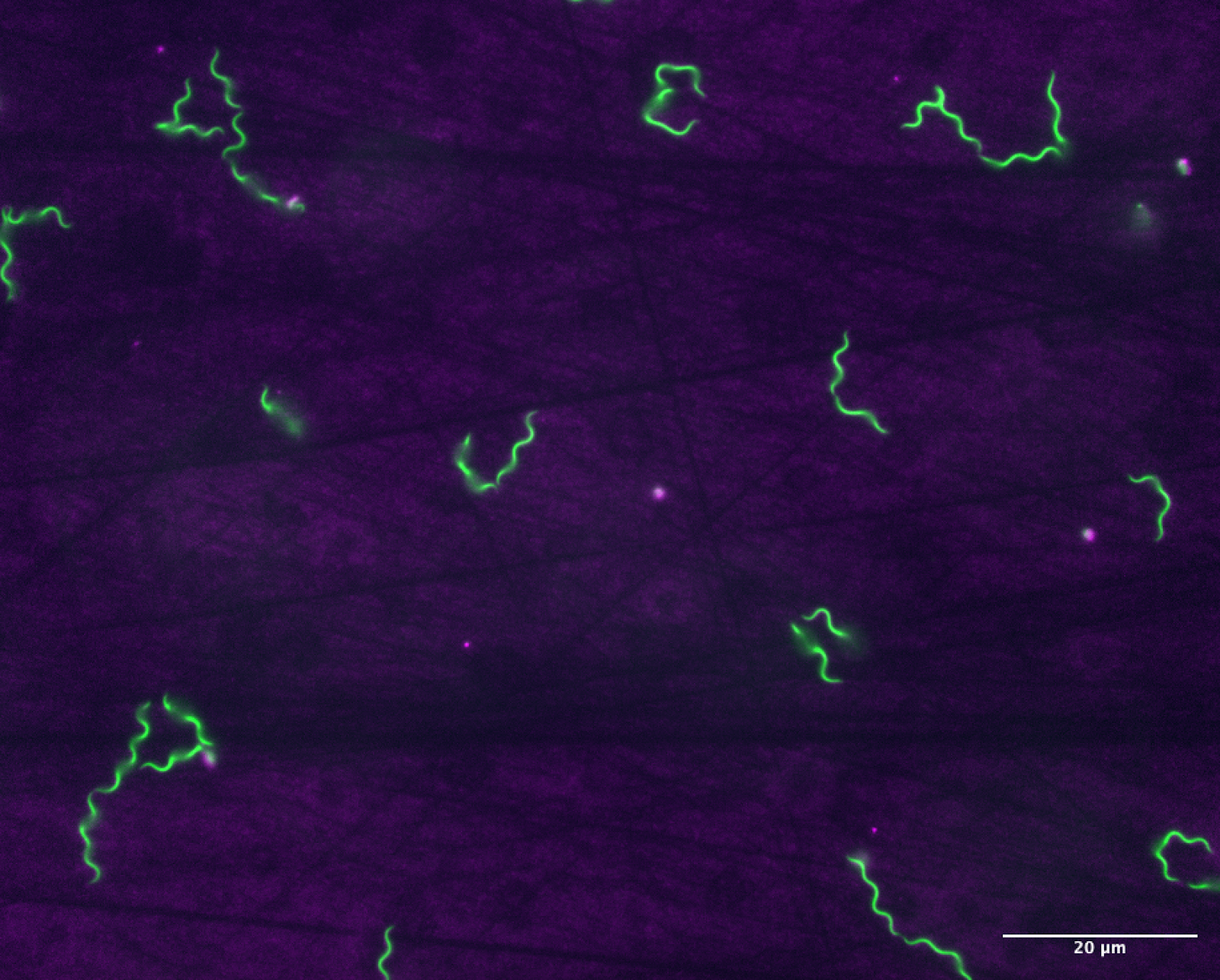
Spirochaetes. Photo credit: Malique Bowen (MD 2022 student)
Microbial eukaryotes are cute but mighty

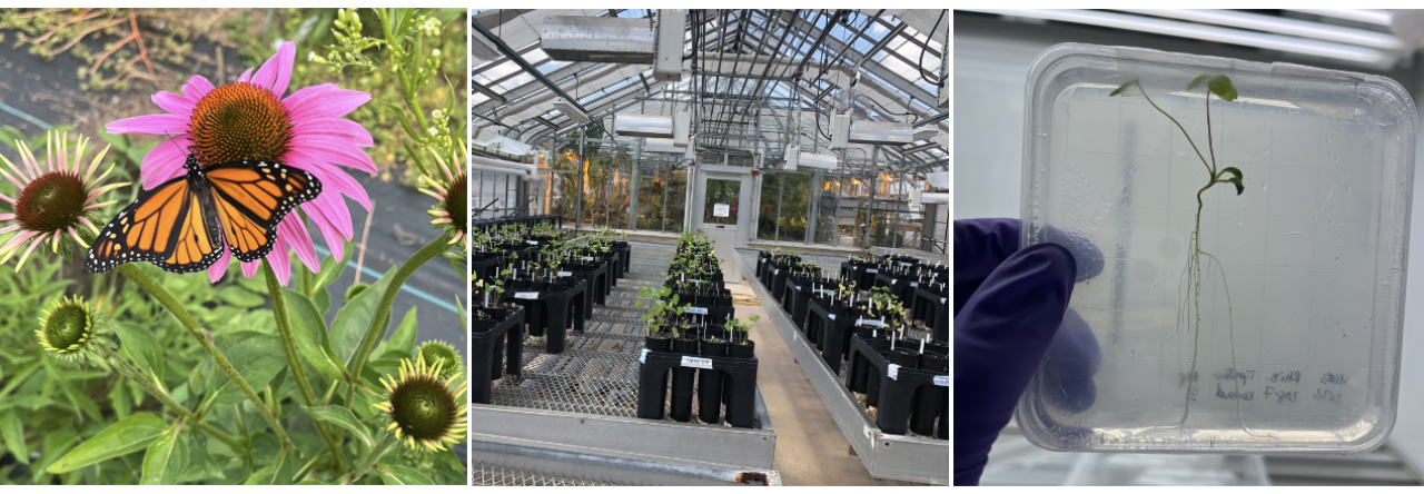
In the last two weeks of the course, we completed individual ‘mini’ projects to research a scientific question of personal interest within Microbial Diversity. For my project, I wanted to work with a group of aquatic microbial eukaryotes called ciliates because I found it fascinating how different they could look and swim, and also they are pretty cute. When the father of microbiology, Antione van Leeuwenhoek, called microbes ‘animalcules’ meaning ‘little animals’, he was pretty spot on in the case of ciliates. So for my research question, I was interested in determining how tolerant different types of ciliates were to relevant environmental stressors: copper, lithium, and sulfur. Copper and lithium are common sources of heavy metal pollution in aquatic ecosystems. For instance, copper is often leached into water from antifouling paint used on boats, and lithium is used in batteries to power many electronics, including electric vehicles. Additionally, ciliates that inhabit unique places like the Great Sippewissett Marsh live in highly sulfuric environments, but it’s unknown what range of sulfur they are tolerant to or if there is a limit to their sulfur tolerance. To perform these tests, I recorded movies under the microscope of four different types of ciliates that I isolated from the Great Sippewissett Marsh after they had been exposed to different concentrations of each stressor for eight hours. Then, I used the computer software Fiji to analyze the movies and measure how ciliate swimming speed was impacted by each condition. I expected that ciliate speed would slow down as the concentration of each stressor increased, and the tolerance limit of each ciliate could be identified by the concentration that completely inhibited movement. Ultimately, the timeline for the project was too short for me to come up with conclusive answers, but I did find some evidence that the ciliates differed in their tolerance to copper and lithium. Most surprising was the finding that one ciliate type could withstand 63 mg/L of copper (II) sulfate, which is a level of copper that not only killed all other ciliates but also totally changed their shapes. Since ciliates are important food sources at the bottom of aquatic food webs, it would be interesting to see how this striking variation in copper tolerance would impact other organisms that feed on them and the broader ecosystem.


There is still so much to discover!
Although the Microbial Diversity course taught me so much about the different microorganisms that have been found in nature, it also taught me that there are way, way more microorganisms that we do NOT know about. Actually, the numbers are mind-boggling. In the ocean, there are 100 million times as many bacteria as there are stars in the known universe. If you picked up a teaspoon of soil, there would be about 1 x 10⁹ bacteria, which is roughly the same number of humans that currently live in the whole continent of Africa. And if all the viruses on earth were laid end to end, they would stretch for 100 million light years (see this Nature article for these astounding estimates and more). Overall, it’s estimated that there are upwards of one trillion microbial species on earth, and we have still to discover 99.999% of them. So, while we are learning more and more that microbes can have strong impacts on human health and the environment, there is still so much that is unknown about microbial diversity. Thanks to the Microbial Diversity course, I feel much more confident in my abilities as a plant biologist turned microbiologist and look forward to making new discoveries in this microbial world of ours.